
 Nana Banana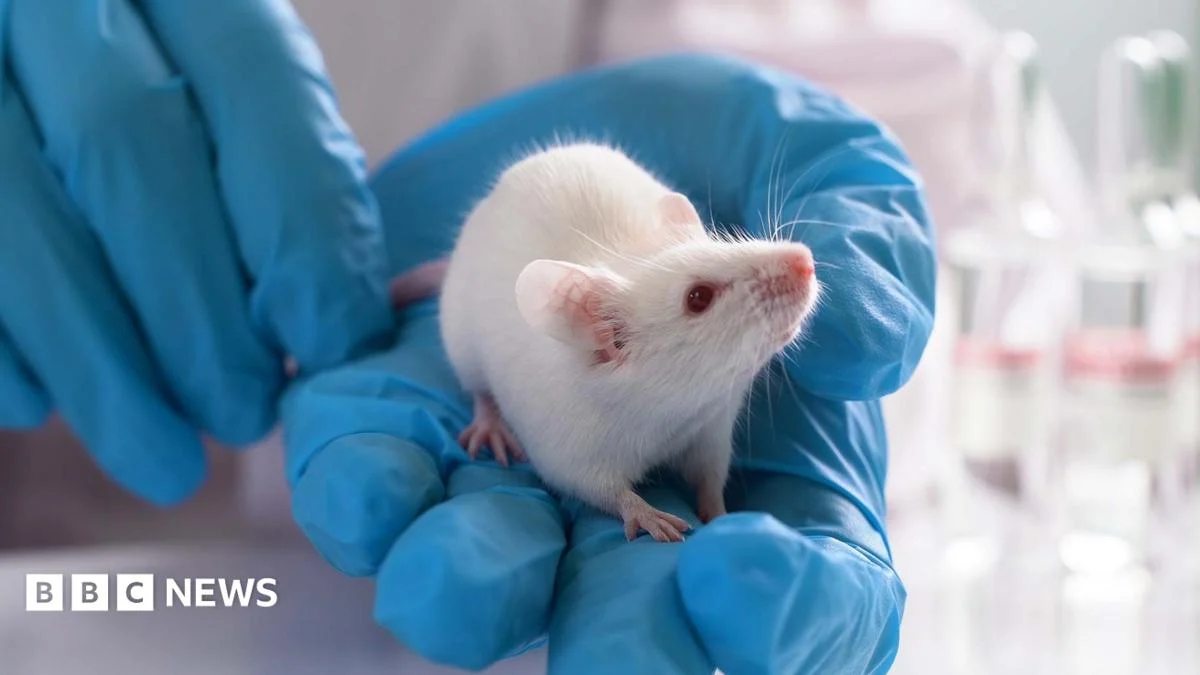
This groundbreaking and ethically complex scientific advancement aims to revolutionize the understanding of brain disorders, many of which currently lack effective treatments. The profound challenges in neurological and psychiatric research stem partly from the limitations of traditional animal models. Conditions such as autism spectrum disorders, schizophrenia, and certain forms of epilepsy manifest uniquely in humans, with rodents often failing to develop analogous pathologies or exhibit the intricate cognitive and behavioral deficits seen in human patients. This biological disparity has created a significant hurdle in translating promising laboratory findings into successful clinical interventions.
As Professor Sergiu Pașca, the lead researcher from Stanford University, articulated in a press conference, the field of psychiatry faces "one of the lowest success rates for clinical trials." He highlighted the disheartening reality that "even drugs that actually make it to clinical trial – that seem to be working really well in animal models – fail dramatically in clinic." This consistent pattern of preclinical success followed by clinical failure underscores a critical gap in scientific knowledge: a lack of comprehensive understanding of human brain biology at a fundamental level. Capturing this missing information, Pașca emphasized, is not merely beneficial but "essential" for future therapeutic progress.
The Stanford researchers believe that, for a range of complex neurological conditions including severe epilepsy, various forms of autism, and cerebral palsy, this novel model has the potential to be "transformative." Pașca asserted that "here we have a new model that allows us to actually capture aspects of human brain function in a way that has not been possible before." This breakthrough offers an unprecedented window into the human brain’s development, function, and dysfunction within a living, integrated system.
The human brain is an organ of unparalleled complexity, comprising an estimated 86 billion neurons and trillions of synaptic connections, organized into millions of intricate circuits. This astonishing density and interconnectedness make it exceptionally difficult to decipher its developmental processes and precisely identify the cellular and molecular mechanisms that go awry in disease states. Traditional methods, such as post-mortem analysis or two-dimensional cell cultures, offer limited insight into the dynamic, three-dimensional interactions characteristic of a living brain.
While the implantation of human neurons into laboratory rodents is not an entirely novel concept, previous studies typically involved simpler grafts or less integrated systems. This latest research elevates that approach to an entirely new level of sophistication and functional integration. The scientists initiated their process by genetically engineering mice to suppress the development of almost their entire cerebral cortex. This outer layer of the brain, often referred to as "grey matter," is responsible for higher-level cognitive functions, including abstract thought, complex memory formation, sensory processing, and conscious awareness. By creating mice largely devoid of their native cerebral cortex, the researchers effectively prepared a biological "scaffold" or "incubator" for human brain tissue.
Next, the researchers utilized human skin cells, which were "reprogrammed" using induced pluripotent stem cell (iPSC) technology. This innovative technique allows adult somatic cells to be reverted to an embryonic-like state, from which they can then be directed to differentiate into virtually any cell type in the body – in this case, forming brain-like tissue. These laboratory-grown structures, termed organoids, are not complete, fully functional brains in a dish. Instead, they are intricate, three-dimensional collections of connected, living cells, comprising various neuronal and glial cell types that self-organize to mimic aspects of the developing human brain architecture. They possess a remarkable capacity for spontaneous electrical activity and rudimentary circuit formation, offering a more biologically relevant model than two-dimensional cell cultures.
Upon implantation into the genetically-engineered mouse brains, these human cerebral organoids did not merely survive; they thrived. The human cells exhibited robust division and differentiation, actively organizing themselves into the existing neural circuitry of the animal. Crucially, they established functional connections with the host mouse’s brain and spinal cord, indicating a level of integration that far surpasses previous attempts. Electrophysiological recordings and neural tracing techniques confirmed that the human neurons were not only receiving input from the mouse brain but also sending signals, suggesting active participation in the chimeric brain’s function.
However, the resulting chimeric cortex was not a perfect replica of a normal human or even mouse cortex. Dr. Ilary Allodi, a neuroscientist from St Andrews University who was not involved in the research, noted that scans of these human-mouse brains appeared "a bit messy." This observation refers to the less-organized, less-stratified appearance compared to the highly structured, layered organization characteristic of a naturally developed cerebral cortex. Despite this initial structural imperfection, over several months, the implanted human cells matured, beginning to exhibit both the morphological characteristics and electrophysiological functions expected of the outer layer of a developing brain.
Approximately six months after the surgical implantation, the scientists subjected the mice to a series of basic behavioral tests. These tests involved observing the mice’s movements and interactions within a small, controlled table-top arena, designed to assess general motor function, exploratory behavior, and basic sensory processing. Professor Pașca reported that the mice with human brain tissue performed "largely as [the normal] mice did," exhibiting no discernible cognitive or behavioral enhancements. This finding is significant for ethical considerations, as it suggests the human brain tissue did not confer human-like intelligence or consciousness upon the mice.
Dr. Sarah Chan, a reader in bioethics at the University of Edinburgh, who was not involved in the study, weighed in on the profound ethical implications. She reassured that there was "no indication that what’s being created here are mice that can think like humans, or a human brain in a mouse body." Nevertheless, she highlighted that the study "prompts us to think about what it might mean when we start changing animal cognition." Dr. Chan posed critical questions: "How can we know what it’s like to be one of these mice? And how do we take account of that in the ways that we treat laboratory animals?" These questions underscore the ongoing debate about the moral status of chimeric organisms and the responsibilities researchers bear when manipulating fundamental biological identities.
Dr. Allodi praised the technical prowess of the scientists, describing their work as "very impressive." She particularly emphasized the remarkable discovery that specific cell types, typically found exclusively in human and other primate brains—and absent in rodent brains—spontaneously formed within the implanted human organoids in the mouse environment. These human-specific cells, such as certain types of interneurons or uniquely complex astrocytes, are thought to play crucial roles in higher-order cognitive functions and may be implicated in the unique pathologies of human brain disorders. Allodi eloquently summarized this phenomenon by stating, "You’re keeping the human program inside the mouse environment — like the mouse is an incubator." This in vivo environment provides essential advantages over in vitro cultures, including a robust blood supply, complex neurotrophic support, and integration into active neural networks, facilitating the long-term survival and maturation of the human tissue.
The researchers confirmed that these chimeric mice were developed and reared under stringent ethical and welfare guidelines, adhering to established protocols for animal research. Due to the inherent ethical complexities, technical demands, and high resource investment, these sophisticated models are expected to be utilized in a limited number of specialized laboratories. Their application will likely be focused on studies of a select few, very specific brain disorders where human-specific biological insights are paramount.
Professor James Ainge, also a neuroscientist at St Andrews University, acknowledged the technical brilliance of the development but cautioned that these mice might be "of limited use" in broader research contexts. He reiterated that this limitation stems partly from the "ethical issues of raising living human brain tissue in a mouse and what that would mean for the experience of the animal." Beyond ethics, practical considerations such as the high cost of production, the specialized expertise required for their maintenance, and the inherent variability in organoid development may also restrict their widespread adoption.
Despite these challenges and ethical considerations, Dr. Allodi highlighted the overarching struggle faced by neuroscientists attempting to decipher human brain diseases. "We try to understand human disease, and what we have is mice, cells that live on a plate, and neural networks on a computer," she observed. Each of these existing models offers only a partial glimpse into the complexities of human neurological conditions. This new development, by providing a living, integrated system that incorporates human-specific brain tissue, represents a crucial "new avenue" in brain research. It offers an unprecedented bridge between simplistic in vitro models and the intractable complexity of the human brain, promising to unlock new insights into disorders that have long eluded effective treatment.



 atisusanti
 George Anjay
 Nakalama John

 Ramos Leo